Extraction, Isolation, Characterization and Nootropic Activity of Bioactive Compounds from Ethanolic Extract of Leaves of Leucaena leucocephala
Dinesh Kumar1* and Asheesh Kumar Gupta1, Navneet Verma2 and Sushil Kumar1
1School of Pharmaceutical Sciences, IFTM University, Lodhipur-Rajput, Delhi Road, Moradabad, Uttar Pradesh, India.
2Pharmacy Academy, IFTM University, Lodhipur Rajput, Delhi Road, Moradabad, Uttar Pradesh, India.
Corresponding Author E-mail: dineshpharma181@gmail.com
DOI : http://dx.doi.org/10.13005/ojc/400426
Article Received on : 20 Mar 2024
Article Accepted on : 16 Aug 2024
Article Published : 29 Aug 2024
Reviewed by: Dr. Rakshit Pathak
Second Review by: Dr. Narendra Bhojak
Final Approval by: Dr. B. K Sharma
The current work aims to screen out the phytochemical screening of extract of ethanol extracted from dried leaves of Leucaena leucocephala. Specifically, the soxhlet extraction process was used, along with phytochemical analysis, separations, and the isolation of biologically active compounds using Thin Layer Chromatography (TLC), column chromatography and HPTLC, respectively. The isolated biologically active compound was then characterized using a variety of spectral analysis namely Infra-Red (IR), 1H nuclear magnetic resonance (1HNMR), 13C nuclear magnetic resonance (13CNMR), and mass spectroscopy. Phytochemicals such as carbohydrates, alkaloids, saponins, tannins, flavonoids, lipids, fixed oils, and phenols are found in the plant's ethanolic leaf extract. Spectroscopic methods were used to characterize the single isolated compound, and the findings presented that the structure of isolated compound LLQ was quercetin. Nootropic action was demonstrated by the secondary metabolites present in the ethanolic extract.
KEYWORDS:Leucaena leucocephala; Nootropic activity; Phytochemical analysis; Quercetin
Download this article as:| Copy the following to cite this article: Kumar D, Gupta A. K, Verma N, Kumar S. Extraction, Isolation, Characterization and Nootropic Activity of Bioactive Compounds from Ethanolic Extract of Leaves of Leucaena leucocephala. Orient J Chem 2024;40(4). |
| Copy the following to cite this URL: Kumar D, Gupta A. K, Verma N, Kumar S. Extraction, Isolation, Characterization and Nootropic Activity of Bioactive Compounds from Ethanolic Extract of Leaves of Leucaena leucocephala. Orient J Chem 2024;40(4). Available from: https://bit.ly/3X3icLQ |
Introduction
Alzheimer’s disease (AD) is the very popular and well-known neurodegenerative illness associated to ageing those results in death. It is characterized by a progressive loss of brain function, cholinergic function deterioration, and memory impairment. AD is one of the most frequent kinds of dementia that affect the older population globally1. In 2019 world Alzimer’s Reportpresented that Nearly 50 million individuals worldwide suffer from dementia. By 2050, there will be 152 million of them, doubling every 20 years2.Memory loss, cognitive decline, behavioral abnormalities, and psychiatric disorders are just a few of the symptoms that might be associated with the AD3.The principal medications applied in clinical practice today to treat AD are acetylcholinesterase blockers and N-methyl-D-aspartate (NMDA) receptor blockers4.
Leucaena leucocephala (Lam.) de Wit is a famous fodder plant from the Fabaceae (Leguminosae) family that is widely used in Tamil Nadu, India. However, this plant has excreted its therapeutic qualities. This plant showed the medicinal importance due to the presence of various secondary metabolites. It is an average height, fast developing tropical plant that has expanded rapidly into many tropical and sub-tropical countries5. The leaf, flower, root, and bark of Leucaena leucocephala offer significant therapeutic potential for treating a variety of diseases including antifungal activity6, anthelmintic activity7, anticancer activity8, antioxidant activity9, antidiabetic activity10.
The key goal of this research work was to assess the possible anti-AD properties of the chemical components of Leucaena leucocephala ethanolic leaf extract.
Materials and Methods
Chemicals and Instruments
No purification was done before using any chemicals or solvents of the analytical grade. A Soxhlet apparatus was used for the extraction of leaves. To the separation and isolation of the chemical constituent, Thin Layer Chromatography and column chromatography was used. IR, 1HNMR, 13CNMR and mass spectroscopy was utilized to characterize the chemical constituents.
Plant Collection and authentication
To prepare the ethanolic extract, fresh, healthy leaves of Leucaena leucocephala were procured from Vill Karanpur Mafi post Hasanpur Distt Amroha. The plant material has been submitted as a voucher specimen in the Herbarium of NISCAIR, New Delhi, India. NIScPR/RHMD/Consult/2021/ 3914-15-2 was the authentication number.
Extraction
Leucaena leucocephala fresh leaves were air dried for about two weeks at room temperature in the shade. The leaves were powdered once they had thoroughly dried. The extraction process employed a Soxhlet extraction apparatus. The leaf powder was extracted with Petroleum ether (60-80°C) for defatted and after this extraction was made with ethanol. Within the apparatus’s upper chamber, a porous thimble held one hundred g of powdered leaf material. To the lower boiling flask, two hundred g of extracting solvent were added. A thermostat-controlled heating mantle was used to heat the flask. The round-bottom flask was filled with ethanol, and the solvent’s boiling point was used to determine the temperature. After heating the solvent to reflux, it was extracted. Up till a dark green extract was gathered on top of the extractor, the material in the thimble was extracted one at a time. Following collection, the ethanolic extract was concentrated independently under low pressure. Following the extraction of ethanol, the extract was weighed and stored at 5°C in a brown, sealed bottle until needed.
Phytochemical Analysis
The ethanolic extract underwent phytochemical examination to identify various components active secondary metabolites or components like tannins, alkaloids, flavonoids, terpenoids, steroids, carbohydrates, proteins, and saponins11.
Isolation and characterization
Thin Layer Chromatography
TLC experiment was carried out on pre-coated Merck silica gel 60 PF254 aluminium sheets, and column chromatographic separation was executed with silica gel (60–120 mesh). Iodine vapors helped to visualize the spots. Toluene, ethyl acetate, chloroform, and Glacial acetic acid were used as mobile phase in ratio 4:3:2:1.
HPTLC Analysis for quantitative measurement of Catechin and Quercetin
Additionally, we used HPTLC to determine the quantity of catechin and quercetin in leaf extract of Leucaena leucocephala. Since HPTLC is a straightforward and less expensive method than other method, we used it for quantitative estimation.
Table 1: Solvent system used in HPTLC.
|
Plant |
Solvent System |
Ratio |
Detection |
|
Leucaena leucocephala |
Toluene: Ethyl Acetate: Chloroform: Glacial Acetic Acid |
4: 3: 2: 1 |
UV254 |
Preparation of Reference solution
Reference solution having a con. of 1000 µg/ml were prepared by mixing standard doses of quercetin and catechin, in alcohol with 100 mg.
Preparation of test solution
Hundred milligrams of ethanolic extract obtained from Leucaena leucocephla was dissolved with 10 ml C2H5OH, mixed properly and then filtered. After the filtration process, filtrate was utilized to perform the HPTLC analysis.
Method Development for HPTLC
HPTLC is most popular techniques to figuring out how much phytoconstituent is present in different parts. Using a Camag Linomat V, an aliquot of each diluted solution (reference and test) was put on a pre-coated TLC plate. The volume of the aliquot was ten µl. The chromatogram was prepared in the presence of saturated chamber using the solvent system listed above, which is also indicated in Table 1. After being dried with a hot air stream, the formed plate was analyzed in a TLC scanner at a wavelength that could be detected. The peak area and area under the curve were used to draw a calibration curve against the concentrations of sample. Using an equation of regression based on the calibration curves, the amount of the marker compounds was calculated12.
Characterization
The melting point was observed using the open capillary tube method, which is the standard method. Uncorrected results were noted for the findings. Using a Bruker Avance Neo 500 MHz spectrometer in DMSO, the 1HNMR spectra were captured. The internal standard that was employed was Tetramethyl silane (TMS). The 1HNMR chemical shifts were measured from TMS downfield in parts per million (ppm). Utilizing a Bruker Avance Neo 500 MHz spectrometer in DMSO, the 13CNMR spectra was captured. The mass spectrum was recorded using a mass spectrometer. Using spectroscopic methods, the structures of the chemical derived from the ethanolic extract were clarified13.
Biological activity
Animals used in Experiment
Albino rats (140–200 g) subjected in biological research. The Albino rats were acquired from the IFTM University’s Institutional Animal House in Moradabad, Uttar Pradesh. Before trial began, the animals spent seven days in a lab setting. The rats were reserved at room temperature (RT) of 24±1°C, with a light and dark cycle (12h/12h). The rats were randomly assigned to positive, negative and test groups during the acclimation period. The animals were kept apart on sterile rice husk bedding in polyacrylic cages. Every animal received an ad libitum supply of fresh water and a normal pellet meal. The Animal Ethical Committee, CPCSEA, Government of India, examined and approved the protocols for animal-based experiments. The clearance number for these protocols is 2021/837ac/PhD/02.
Acute Toxicity
Using female Wistar albino rats, the acute toxicity of an ethanolic extract was assessed. Acute toxicity was performed according to OECD guideline 423. 2 groups of three animals each were used for the experiment. Before the trial, they fasted for 12 hours while having unrestricted access to water. One group received a single oral dosage of 2000 mg/kg of ethanolic extract, whereas the other received normal saline as treatment. After administering the ethanolic extract, the treated animals were monitored continuously for 30 minutes to check for any changes in overall behavior, water and food intake, and death. Thereafter, they were monitored sporadically for 4 to 24 hours. The rats were monitored again for a maximum of 14 days after the treatment14.
Biological Evaluation
Nootropic Activity
Nootropic activity evaluation was done by using Y Maze and Novel objective recognition test (NORT).
NORT
The rats were arranged in six groups and every group consists of 6 rats and presented as
Group 1: Normal control Group 2: Scopolamine (Scop) 1mg/kg/day Group 3: Scop + EEAC 100mg/kg/day Group 4: Scop (1mg/kg/day) + EEAC 200mg/kg/day, Group 5: Scop (1mg/kg/day) + EEAC 400mg/kg/day Group 6: Scop (1mg/kg/day) + Donepezil (2 mg/kg/day). Treatment was given to rats for 21 days.
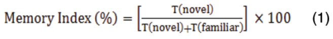
Apparatus was a white-coloured open box of plywood. The box length was l70 cm, wide was 50 cm and height were 40 cm. Experiments carried out in three phases: habituation, familiarization, and testing. To habituate and diminish the rats’ dread of a new environment, they were exposed to the device without items for 5 minutes, with the freedom to explore the equipment. The rats were placed in the same setting the next day, but with two identical items A1 and A2 (plastic balls). The familiarization exploring time was 5 minutes. After 24 hours the rats were introduced for testing phase. During this phase, rats were returned to the apparatus, which contained a novel item (Plastic Square) utilized to modify A2. When the rat smelled or made contact with the things with their nose, this was termed exploration. The contact time for plastic square and the plastic ball were noted. To hide any smell residues, the device was washed with disinfectant at the end of each session. The discrimination index/Memory Index (DI) was calculated by using below formula 1 15.
Y Maze
Animals were grouped as per the NORT task. This apparatus employed to estimate percentage alteration (Memory Index) of the experimental rats. Y maze used in this experiment contains three similar arms, P, Q and R. The arm was 40 centimeter in length, 35 cm in height and 12 cm in wirth, positioned at 120ᵒ angles. For the test, every animal was sited on maze triangle center and permitted to freely explore it for 8 minutes. The arm entry was pronounced complete if all the body part completely in the arm. Series of arm entries was visually recorded. To avoid odors, the maze floor was washed with 70% ethanol between trials. The overall number of visits was utilized to calculate the degree of percentage alternation (memory index) using the below formula 216.

Results and Discussion
Percentage extraction yield and physical properties of ethanolic extract
Extraction of leaves was done by using ethanol. It was obtained that the ethanolic extract’s percentage extraction yield was 8.36% w/w. The ethanolic extract demonstrated dark brownish colour. The consistency of ethanolic extract was found to be sticky.
Preliminary phytochemical screening
A standard process was used to check for phytochemicals. Extracted ethanolic extract was utilized for the preliminary phytochemical screening and demonstrated the attendance of carbohydrates, alkaloids, saponins, tannins, flavonoids, and phenols and showed in table 2.
Table 2: Findings of Phytochemical screening
|
Sr. No. |
Phytoconstituent Class |
Test Name |
Results |
|
1 |
Carbohydrates |
Fehling’s |
+ |
|
2 |
Alkaloids |
Dragendroff’s |
+ |
|
3 |
Saponins |
Foam Test |
+ |
|
4 |
Test for Steroids |
Salkowski Reaction |
– |
|
5 |
Tannins |
Ferric Chloride test |
+ |
|
6 |
Tests for Glycosides |
Borntrager’s Test |
– |
|
7 |
Flavonoids |
Shinoda test |
+ |
|
8 |
Test for Fats and Fixed oils |
CuSO4 |
– |
|
9 |
Test for Phenol |
Ferric chloride test |
+ |
Thin Layer chromatography and Column Chromatography
Using the solvents Toluene, Ethyl Acetate, Chloroform, and Glacial Acetic Acid in the proportions 4:3:2:1 (v/v), a TLC finger print profile was established. Using an iodine chamber, five spots were seen (Fig. 1). Compound LLQ was the only one of the five that could be satisfactorily extracted using column chromatography and eluted with n-hexane and chloroform.
 |
Figure 1: Pictogram of developed TLC plate of ethanolic extract of Leucaena leucocephala |
HPTLC Analysis
The results of HPTLC analysis exhibited the presence of catechin and quercetin in the ethanolic leaves extract of Leucaena leucocephala (Figure 2 and 4). Chromatogram of Marker compound showed in figure 5. Rf value of catechin and quercetin of ethanolic leaves extract of Leucaena leucocephala was similar to the marker compounds at 0.17 and 0.47 when the plates were scanned at 254nm and showed in table 3. The calibration curve of catechin and quercetin was prepared and correlation coefficient was 0.99 presented in figure 3A and 3B. The quantity of catechin and quercetin in the ethanolic leaves extract of Leucaena leucocephala was 392.16µg and 619.72µg /100 mg.
Table 3: Spot no and Rf values of marker compounds and ethanolic extract of Leucaena leucocephala at UV 254nm.
|
Spot No |
Marker Compounds (Rf) |
Leucaena leucocephala (Rf) |
|
1 |
0.17(catechin) |
0.11 |
|
2 |
0.47(quercetin) |
0.17 |
|
3 |
0.32 |
|
|
4 |
0.47 |
|
|
5 |
0.52 |
|
|
6 |
0.64 |
|
|
7 |
0.72 |
|
|
8 |
0.84 |
|
|
9 |
0.90 |
 |
Figure 2: HPTLC Finger print of ethanolic extract of Leucaena leucocephala scanned at 254nm. |
 |
Figure 3A: Calibration curve of Catechin |
 |
Figure 3B: Calibration curve of Quercetin. |
 |
Figure 4: Chromatogram of Leucaena leucocephala scanned at 254nm |
 |
Figure 5: Chromatogram of standard Catechin and Quercetin scanned at 254 nm |
Characterization
Characterization of isolated compound was established using IR, 1H NMR, 13C NMR and Mass spectroscopy and spectral data are presented below
Colour: Yellow, M.P: 314-16 ᵒC, IR (KBr) cm-1: 1517 (C-C str.),1467 (C=C str.), 2997 (C-H str.), 1649 (C=O str.), 1319 (C-O str.), 1612 (O-H str.);1HNMR (500MH2, DMSO-d6): 6.40-7.68- (5H, M, Aromatic); 5.05-5.52- (5H, S, OH); 13CNMR (500MH2, DMSO-d6): 93.45, 98.28, 103.09, 115.15, 115.69, 120.10, 122.07, 135.80, 145.11, 146.86, 147.75, 156.22, 160.78, 163.96, 175.89; MS (FAB) [M + 1]+m/z): 303.02
Compound LLQ was identified as Quercetin by comparison of physical properties and spectral data with previously reported data.
 |
Scheme 1 |
Oral Acute Toxicity Study
During the 14d observation cycle in the investigation of acute toxicity. It was found that no toxic effect and no death was occurred after the administration of single dose upto 2000mg/kg orally. There were no discernible alterations in the body weight increases. Therefore, Leucaena leucocephala safe in doses up to 2000 mg/kg of animal weight. Table 4 displays the parameters that were obtained for the acute toxicity research following the administration of the ethanolic extract in comparison to the normal group.
Table 4: General appearance and behavioral symptoms during the acute toxicity study.
|
Observation |
Control Group |
Treated Group (2000 mg/kg) |
|
Body weight |
– |
– |
|
Body Temperature |
– |
– |
|
Intake of food |
– |
– |
|
Urination |
– |
– |
|
Respiration rate |
– |
– |
|
Change in skin |
– |
– |
|
Eye color |
– |
– |
|
Sedation |
– |
– |
|
Drowsiness |
– |
– |
|
Diarrhea |
– |
– |
|
Coma |
– |
– |
|
Death |
– |
– |
+ = Present, – = Absent
Nootropic Activity
Novel object recognition test (NORT)
NORT results are exhibits in figure 6. Figure 6 depicts the percentage memory index on the NORT apparatus across the treatment groups. In the scop group the % memory index was considerably less than the control group (p<0.05). EELL in a dose 100 mg/kg was not exhibited the potent action but dose 200 and 400mg/kg produce significant effect in dose dependent manner and Donepezil 2 mg/kg considerably increased % memory index to the Scop group (p < 0.05). These findings showed that EELL in all dose prevented memory deficits produced by Scop in a dose-dependent way.
 |
Figure 6: Effect of EELL on NORT in Scop induced memory impaired rats. Data expressed as mean ± SEM (n=6). *p<0.05 vs control group, # <0.05 vs Scop group. |
Y Maze
Y maze results are exhibits in figure 7. Figure 7 depicts the percentage memory index on the Y maze across the treatment groups. In the scop group the % memory index was considerably less than the control group (p<0.05). EELL in a dose 100 mg/kg was not showed the significant anti Alzheimer’s effect but dose 200 and 400mg/kg produce significant effect in dose dependent manner and Donepezil 2 mg/kg considerably increased % memory index to the Scop group (p < 0.05). These findings showed that EELL in all dose prevented memory deficits produced by Scop in a dose-dependent way.
 |
Figrue 7: Effect of EEAC on memory index on Y Maze in Scop induced memory impaired rats. Data represented as mean ± SEM (n=6). *p<0.05 vs control group, #<0.05 vs Scop group. |
Conclusion
8.36% w/w percentage yield was obtained from ethanolic extract. A number of phytochemicals namely carbohydrates, saponins, alkaloids, tannins, flavonoids, and phenols was found to be present in the ethanolic extract of leave. In acute toxicity study, no toxic effect was found. The results suggested that secondary metabolites were responsible for nootropic activity. HPTLC was used to determine the quantities of marker compounds. The isolation was done from ethanolic extract and spectroscopic methods were used to characterize the isolated compound which demonstrated the chemical structure of compound LLQ as Quercetin.
Acknowledgement
This research work is the part of Doctor of Philosophy degree from IFTM University, Lodhipur-Rajput, Moradabad. Authors want to say the thanks for the administration of IFTM University for providing the facilities.
Conflict of interest
There is no conflict of interest.
References
- Kamel, N.N.; Aly, H.F.; Fouad, G.I.; El-Karim, S.S.A.; Anwar, M.M.; Syam, Y.M.; Elseginy, S.A.; Ahmed, K.A.; Booles, H.F.; Shalaby, M.B.; Maha, Z. Rizk. Anti-Alzheimer activity of new coumarin-based derivatives targeting acetylcholinesterase inhibition. RSC Adv. 2023; 13(27): 18496–18510.
CrossRef - Subash, P.; Kareti, S.R.In silico molecular docking analysis for potential anti-Alzheimer’s compounds from the methanolic leaf extract of Erythroxylum monogynum using Gas chromatography–mass spectrometry. J. Saudi Chem. Soc. 2021; 25(8): 101285.
CrossRef - Simunkova, M.; Alwasel, S.H.; Alhazza, I.M. Management of oxidative stress and other pathologies in Alzheimer’s disease. Arch. Toxicol 2019; 93: 2491–513.
CrossRef - Song, X.; Wang, T.; Guo, L.; Jin, Y.; Wang, J.; Yin, G.; Jiang, K.; Wang, L.; Huang, H.; Long L. In Vitro and In Vivo Anti-AChE and Antioxidative Effects of Schisandra chinensis Extract: A Potential Candidate for Alzheimer’s Disease. Evid. Based Complement Alternat Med. 2020; 2020: 2804849.
CrossRef - Deivasigamani, R. Phytochemical analysis of Leucaena leucocephala on various extracts. J. Phytopharmacol. 2018; 7(6): 480-482.
CrossRef - Elbanoby, N.; EEl-Settawy, A.A.A.; Mohamed, A.A.; Salem, M.Z.M. Phytochemicals derived from Leucaena leucocephala (Lam.) de Wit (Fabaceae) biomass and their antimicrobial and antioxidant activities: HPLC analysis of extracts. Biomass Conver. Biorefnery. 2022,
CrossRef - Widaad, A.; Zulkipli, I.N.; Petalcorin, M.I.R. Anthelmintic Effect of Leucaena leucocephala Extract and Its Active Compound, Mimosine, on Vital Behavioral Activities in Caenorhabditis elegans. Molecules. 2022; 27(6): 1875.
CrossRef - Zarin, M.A.; Wan, H.Y.; Isha, A.; Armania, N. Antioxidant, antimicrobial and cytotoxic potential of condensed tannins from Leucaena leucocephala hybrid-Rendang. Food Sci. Human Wellness. 2016; 5(2): 65-75.
CrossRef - Benjakul, S.; Kittiphattanabawon, P.; Sumpavapol, P.; Maqsood, S. Antioxidant activities of lead (Leucaena leucocephala) seed as affected by extraction solvent, prior dechlorophyllisation and drying methods. J Food Sci Technol. 2014; 51(11): 3026–3037.
CrossRef - Chowtivannakul, P.; Srichaikul, B.; Talubmook, C. Antidiabetic and antioxidant activities of seed extract from Leucaena leucocephala (Lam.) de Wit. Agricul. Natural Resour. Volume 2016; 50(5): 357-361.
CrossRef - Kokate CK. Practical Pharmacognosy. 4th ed. New Delhi: Vallabh Prakashan; 1999.
- Shanmugam M, Subramanian S, Ramachandran S. Method development and validation for quantification of six bioactive compounds (andrographolide, columbin, piperine, gallic, paracoumaric and oleanolic acids) by HPTLC. J. Complement. Integr. Med. 2023;20(1): 137-145.
CrossRef - Arora, S.; Itankar, P. Extraction, isolation and identification of flavonoid from Chenopodium album aerial parts. J. Tradit. Complement. Med. 2018; 8 (4):476–482.
CrossRef - OECD 423 – Guidelines for the testing of chemicals Acute oral toxicity –Fixed dose procedure. Animals. 2001:1–14.
- Kheradmand, E.; Moghaddam, A.H.; Zare, M. Neuroprotective effect of hesperetin and nano-hesperetin on recognition memory impairment and the elevated oxygen stress in rat model of Alzheimer’s disease. Biomed. Pharmacoth. 2018; 97:1096-1101.
CrossRef - Nazir, N.; Zahoor, M.; Nisar, M.; Karim, N.; Latif, A.; Ahmad, S.; Uddin, Z. Evaluation of neuroprotective and anti-amnesic effects of Elaeagnus umbellataThunb. On scopolamine-induced memory impairment in mice. BMC Complement Med. Thera. 2020; 12: 143.
CrossRef

This work is licensed under a Creative Commons Attribution 4.0 International License.
About The Author
![]()
A New Edition of Web of Science
Journal Impact Factor
2022: 0.5
Five Year: 0.8
Journal is Indexed in

Cabells Whitelist
![]()


