Potential Bactericidal Effect of Silver Nanoparticles Synthesised from Enterococcus Species
Kelmani Chandrakanth R1*, Ashajyothi C1, Ajay Kumar Oli2, Prabhurajeshwar C1
1*Department of Biotechnology, Gulbarga University, Gulbarga-585106, Karnataka, India.
DOI : http://dx.doi.org/10.13005/ojc/300341
Article Received on :
Article Accepted on :
Article Published : 21 Aug 2014
The present study proposed a synthesis of silver nanoparticles (AgNPs) from Enterococcus species. Silver has a strong antimicrobial potential, which has been used since the ancient times. The present work is carried out to screen bactericidal potential of silver nanoparticles against clinically isolated multidrug resistant bacteria’s. Biosynthesized silver nanoparticles were characterized by analytical techniques including as UV-Visible spectrophotometer, Field Emission Scanning Electron Microscopy (FeSEM), Energy dispersive x-ray spectroscopy (EDX), Nanoparticle Tracking Analyzer (NTA) analysis. Antimicrobial effect of silver nanoparticles against E. coli and K. pneumonia and Methicillin resistant Staphylococcus aureus (MRSA) respectively were investigated by Agar well diffusion method.
KEYWORDS:Silver nanoparticles; Enterococcus species; Minimal Inhibitory Concentration; Bactericidal potential
Download this article as:| Copy the following to cite this article: Chandrakanth R K, Ashajyothi C, Oli A. K, Prabhurajeshwar C. Potential Bactericidal Effect of Silver Nanoparticles Synthesised from Enterococcus Species. Orient J Chem 2014;30(3). |
| Copy the following to cite this URL: Chandrakanth R K, Ashajyothi C, Oli A. K, Prabhurajeshwar C. Potential Bactericidal Effect of Silver Nanoparticles Synthesised from Enterococcus Species. Orient J Chem 2014;30(3). Orient J Chem 2014;30(3). Available from: http://www.orientjchem.org/?p=4263 |
INTRODUCTION
Before the beginning of antibiotics therapy, silver was used for its antiseptic activity, specifically for the treatment for open wounds and burns (1). Ancient people were much aware of the bactericidal properties of silver. The silver ions are highly reactive, and they bind to proteins followed by structural changes in the bacterial cell wall and nuclear membrane, which leads to cell distortion and death. Silver ions have capacity to inhibit the bacterial replication, by binding and denaturing bacterial DNA (2, 3). Silver ions react with thiol group of proteins, followed by DNA condensation resulting in the cell death (4). Various types of silver compounds that are used as antimicrobials from ancient times include silver nitrate, silver sulfadiazine, silver zeolite, silver powder, silver oxide, silver chloride and silver cadmium powder. Silver nanoparticles have important biological properties as follows: they are effective bactericidal agents against broad spectrum of bacteria, including antibiotic resistant strains (5). The bactericidal activity of silver nanoparticles against the pathogenic, MDR as well as multidrug susceptible strains of bacteria was studied by many scientists, and it was proved that the silver nanoparticles are the powerful weapons aga inst the MDR bacteria such as Pseudomonasaeruginosa, ampicillin-resistant Escherichia coli, erythromycin-resistant Streptococcus pyogenes, methicillin- resistant Staphylococcus aureus (MRSA) and vancomycin-resistant Staphylococcus aureus (VRSA) (6). Bactericidal efficacy of silver nanoparticles was investigated by many researchers and their effective potential against broad range of microbes was proved, including antibiotic-resistant bacteria. Silver nanoparticles are also termed as new-generation of antimicrobials (4). The group of researchers actively proved the bactericidal potential of silver nanoparticles. Feng et al., (6) reported the bactericidal potential of silver ions against S. aureus and E. coli.
Synthesis of the silver nanoparticles by Fusarium acuminatum and its bactericidal efficiency of silver nanoparticles against four human pathogenic bacteria, viz. E. coli, Salmonella typhi, Staphylococcus epidermidis and Staphylococcus aureus, were carried out by Ingle et al., (7). Biosynthesis of silver nanoparticles using bacteria, fungi, and plants are already well-documented (8, 9). Jeevan et al., (10) reported the biosynthesis of silver nanoparticles from P. aeruginosa, and its inhibitory effect on important human pathogens, E. coli and Vibrio cholerae. It is also clear that the bacterium P. aeruginosa can be used to synthesize bioactive silver nanoparticles efficiently using inexpensive substances in an eco-friendly and non toxic manner. The synthesis of silver nanoparticles was achieved using Bacillus megaterium supernatant and 1 mM silver nitrate (11).
In the current study, silver nanoparticles were synthesized from Enterococcus sps. And their antibacterial activity was performed against different clinical pathogens.
MATERIALS AND METHOD
Bacterial Culture
The bacterial strain of Enterococcus faecalis (Non pathogenic) were collected from Medical Biotechnology and Phage Therapy Laboratory (MBPT), Department of Biotechnology, Gulbarga University, Gulbarga. The culture was inoculated on Bile esculin azide agar medium and incubated at 370C for 24hrs for obtained pure culture (12).
Preparation of cell free microbial extract
Luria-Bertani broth were prepared, sterilized and inoculated with fresh Enterococcus faecalis culture. The cultured flasks were incubated at 37oC for 72hrs. After incubation the culture were centrifuged at 10,000 rpm for 10 minute and the supernatant were used for silver nanoparticle synthesis.
Synthesis of silver nanoparticles
The bacterial supernatant was added separately to the reaction vessel containing Silver nitrate (AgNO3) at a concentration of 100 mM (v/v) and control (without the silver nitrate). The reaction was carried out in dark conditions for 24 hours, at 370 C, pH: 7 in rotary shaker with 120 rpm (13).
Characterization of silver nanoparticles:
Synthesized silver nanoparticles were characterized by means visual observation and analytical techniques. After 24 hours of incubation the reaction medium is collected and centrifuged at 10,000 rpm for 10 min, supernatant is further used for UV-Visible spectrum using T90+ UV-VIS spectrophotometer and for Nanoparticle Tracking Analyzer (NTA) pattern analysis, for UV-VIS spectrophotometer scanning the spectra between 300 to 600 nm at room temperature.
The size and shape of nanoparticles was analyzed using Field Emission Scanning Electron Microscopy (FeSEM), after centrifuging the reaction mixture, obtained pellet is washed twice with double distilled water and phosphate saline buffer (pH:7.2), air dry the pellet and then used for FeSEM and EDX analysis. The samples slide were prepared by simple drop coating of the suspension of silver nanoparticles (dissolved in buffer) on a carbon-coated copper grid, by simply dropping a very small amount of the sample on the grid, with excess solution removed using blotting paper. The film on the scanning electron microscopy (FeSEM from Carl Zeiss, UK) grid was then allowed to dry under mercury lamp for 5 minutes. FeSEM instrument is equipped with a Thermo energy dispersive x-ray spectroscopy (EDX) attachment.
Antimicrobial activity of silver nanoparticles against MDR pathogens:
Antimicrobial activity by Agar well diffusion method:
According to Clinical and Laboratory Standards Institute (CLSI) (14) guidelines the antimicrobial activity of Ag nanoparticles was evaluated against E. coli (Clinical MDR pathogen) Klebsiella pneumoniae (Clinical MDR pathogen) and clinical pathogen Methicillin resistant Staphylococcus aureus (MRSA) by modification of the agar well diffusion method. Clinically isolated pathogens were inoculated in Luria-Bertani broth and incubate at 370C for 6hrs. Approximately 108 colony- forming units of 100µl of each microorganism were inoculated on Muller Hinton agar (MHA) plates; Agar wells of 5 mm diameter were prepared with the help of a sterilized stainless steel cork borer. Different concentrations (5, 10, 15, 20, 25 µg/ml) of the nanoparticles and maintain water as control in another well, were loaded on marked wells with the help of micropipette under aseptic conditions and plates were incubated at 37ºC for 18 and 24 hrs. The zone of inhibition was measured using a ruler and expressed in mm (15).
Minimum Inhibitory Concentration (MIC)
The bactericidal activity of silver nanoparticles were determining by Broth dilution method according to CLSI 2000 (14). Bacterial cells were grown in 10 ml LB broth by inoculating 10 µl of 18 hrs culture, supplemented with 2, 4, 6, 8, 10, 12, 14, and 16μg ml-1 of silver nanoparticles. All tubes were incubated at 37°C for 18 hrs and 24 hrs and measure the absorbance at 600 nm. Antimicrobial test compound below the MIC cannot inhibit microbial growth.
RESULTS AND DISCUSSION
E. faecalis culture obtained from MBPT laboratory after inoculation onto the selective media Bile esculin azide agar they appears in black color colonies. In further biosynthesis of silver nanoparticles from test strain the primary conformation of synthesis of silver nanoparticles in the medium was characterized by the changes in color from yellowish white to brown (shown in the Fig: 1). The characteristic surface Plasmon resonance of silver nanoparticles ranges between 380 nm to 450 nm due to excitation of surface Plasmon vibrations and this is responsible for the striking yellow brown color of silver nanoparticles (16).
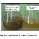 |
Fig1: Visual Observation of Synthesis of Silver Nanoparticles Click here to View Figure |
Biological method of synthesis of silver nanoparticles exhibit strong absorption of electromagnetic waves in the visible range due to their optical resonant property, called Surface Plasmon Resonance (SPR). The characteristic surface plasmon resonance of silver nanoparticles ranges between 300 nm to 325 nm (shown in the Table: 1 and Fig: 2, 3) due to excitation of surface Plasmon vibrations. According to Mie theory, only a single SPR band is expected in the absorption spectra of spherical nanoparticles whereas, the number of peaks increases as anisotropy increases (17). According to Abd El-Raheem et al., (18) report the strong surface of plasmon resonance absorption of silver nanoparticles prepared using E. coli ATCC 8739, B. subtilus ATCC 6633 and S. thermophilus ESh1 was centered at 430, 419, and 420 nm, respectively. In the present study, SPR band reveals spherical shape of silver nanoparticles, which was further conformed by FeSEM and NTA analysis. The spectrum with bands in 300 to 325nm range indicates the presence of nano-sized silver metals in the culture solution. Maximum absorbance is obtained at 309nm (OD: 2.706) for 24hrs of incubation of culture supernatant with 100 mM silver nitrate in (1:1) volume.
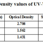 |
Table1: Optical Density values of UV-Visible SpectrumClick here to View table |
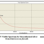 |
Fig2: UV-Visible Spectrum for biosynthesized silver nanoparticles from Enterococcus faecalis Click here to View Figure |
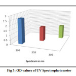 |
Fig3: OD values of UV Spectrophotometer Click here to View Figure |
The diameters of particles were measured to obtain the particle size. Geethalakshmi et al., (19) were reported spherical silver nanoparticles with diameters of 36–94 nm are depicted in the Field emission scanning electron micrographs synthesized from Trianthema decandra (Aizoaceae) root extract and Deshpande et al., (20) also reported 90% of the silver nanoparticles synthesized from Carom Seed (Trachyspermum copticum) extract are in the range of 6 – 50 nm in FeSEM studies. In our present study we are reporting FeSEM images of functionalized silver nanoparticles synthesized from E. faecalis bacterial biomass can be seen with core shell morphology of size 9 – 130 nm and it is observed that there is a marginal variation in the particle size (Fig: 4). Maximum of the particles are in the range of 11-75 nm. The variation in particle size is possibly due to the fact that the nanoparticles are being formed at different times. Higher resolution image at 300 nm shows a group of particles in embedded in an organic moieties making a stable suspension. The particles appear to be polydispersed in nature and are roughly spherical in shape.
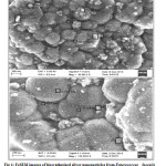 |
Fig4: FeSEM images of biosynthesized silver nanoparticles from Enterococcus faecalisClick here to View Figure |
The study of metallic nature of these silver nanoparticles and reduction of silver into elemental silver and absence of other impurities has been confirmed and further strengthened by EDAX image. The optical absorption peak is observed approximately at 3 keV, which is typical for the absorption of metallic silver nanoparticles due to surface Plasmon resonance (21). In the EDX spectrum of the bacterial mediated silver nanoparticles, the strongest peak detected was from silver with weaker peaks from carbon and oxygen (22). In the current study energy dispersive x-ray spectroscopic profile of silver nanoparticles was confirmed by optical absorption peak at 3 keV, which is typical for silver nanocrystallite absorption. The spectrum shows the nanoparticles are primarily composed of silver with the only noticeable contaminant being phosphorus, sodium elements (Fig: 5).
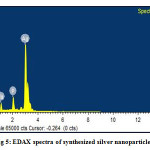 |
Fig5: EDAX spectra of synthesized silver nanoparticlesClick here to View Figure |
Nanoparticle Tracking Analysis is a newly developed method for the direct and real-time visiualization on and analysis of nanoparticles in liquids. According to Pratik et al, (11) report NTA measurements revealed that the mean size of synthesized silver na noparticles was found to be 51 nm with concentration of 5.5 × 10¹⁰ particles/ml in case of bacterial supernatant mediated synthesis. In our studies we revealed that the silver nano sample is quite polydisperse sample with particle size of 152nm and the concentration of sample after dilution comes to 5.6×10¹¹ particles/ml. It is observed that significant numbers of particles are observed from 30 to 300 nm with few aggregates being present at 400-500 nm (Fig: 6, 7 and 8).
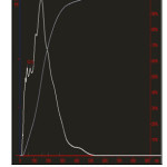 |
Fig6: Particle Size / Concentration of silver nanoparticles Click here to View Figure |
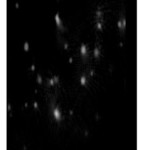 |
Fig7: silver nanoparticle Sample Video Frame Click here to View Figure |
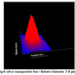 |
Fig8: silver nanoparticle Size / Relative Intensity 3 D plotClick here to View Figure |
The biologically synthesized silver nanoparticles inhibited different pathogenic microorganisms. With the prevalence and increase of microorganisms resistant to multiple antibiotics, silver based antiseptics have been emphasized in recent years. Silver nanoparticles were biosynthesized using fungus Trichoderma viride (23). Ag-NPs have a very broad range of antimicrobial activity and kill both Gram negative and Gram-positive bacteria, including Escherichia coli, Staphylococcus aureus, Bacillus subtilis, Streptococcus mutans and Staphylococcus epidermidis (24, 25, 26, 27, 28, 29, 30, 31).
In the present work antimicrobial activity of biosynthesized silver nanoparticles studied against the multidrug resistant bacteria using standard zone of inhibition. MDR isolate E.coli showed maximum zone of inhibition of 18 mm for 10 µg/ml for K. pneumonia with maximum zone of inhibition of 18 mm for 25μg/ml and Staphylococcus aureus with maximum zone of inhibition of 19 mm for 25μg/ml of silver nanoparticle concentration. The highest antibacterial effect was found with zone of inhibition (19 mm) on Staphylococcus aureus and lowest antibacterial effect in Ag-NPs on Klebsiella pneumonia (14mm). The results were summarized in Table: 2 and Fig: 9, 10 and 11. Among antibiotic, the weakest activity was found to all three MDR pathogenic bacteria’s. In comparison study between antibiotics and silver nanoparticles on E. coli (with antibiotics Rifampicin, Pipracillin, Ceftazidime), K. pneumonia (with antibiotics Ceftazidime, Cephalexin, Ceftriaxone) and Staphylococcus aureus (with antibiotics Ampicillin, Methicillin, Pencillin) showed weakest antibacterial effect as compare to silver nanaoparticles. Results were shown in Table: 3 and Fig: 12.
In Antibacterial activity the resulting zones of inhibition formed were mainly due to the destabilization of the outer membrane, collapse o f the plasma membrane, and depletion of intracellular ATP by the silver nanoparticles (32). 12, 13 and 14 mm zone of inhibition was recorded in E.coli at the concentration of 20, 40, 60μg. In Staphylococcus aureus 11.5, 13.0 and 14.0 mm of zone of inhibition recorded at respective concentrations (33).
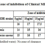 |
Table2: Zone of inhibition of Clinical MDR strains Click here to View table |
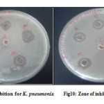 |
Fig9, 10: Zone of inhibition for K. pneumonia, Zone of inhibition for MRSA Click here to View Figure |
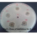 |
Fig11: Zone of inhibition for E. coli Click here to View Figure |
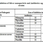 |
Table3: Zone of inhibition of Silver nanoparticle and Antibiotics against Clinical MDR strains
|
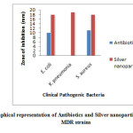 |
Fig12: Graphical representation of Antibiotics and Silver nanoparticles against Clinical MDR strains Click here to View Figure |
The growth of the E. coli cells are inhibited at a concentration of 10 μg/ml of silver nanoparticles. This is the minimum concentration of the nanoparticles which inhibit the growth of the E. coli cells, i.e MIC of Ag nanoparticles (34). Raffin et al., (35) observed that silver nanoparticles with mean sizes of 16 nm were completely cytotoxic for E. coli at a low concentration (60mg/ml). In the present work we report 11μg/ml, 3µg/ml and 12μg/ml of silver nanoparticles was recorded as the minimal inhibitory concentration (MIC) for K. pneumonia, E. coli, Staphylococcus aureus respectively by measuring absorbance at 600 nm shows 0.04 Optical Density indicates the complete inhibition of the growth. As compare to the recently published results, biosynthesized nanoparticles from Enterococcus fecalis showed complete inhibitory action on reported MDR at very low concentrations. Complete inhibitory action of biosynthesized nanoparticle is due to the size of the nanoparticles (9-130 nm), ultrafine particles (size range 10-100 nm) exhibited higher antimicrobial activity than big particles. The antibacterial properties were related to the total surface area of the nanoparticles (36). Smaller particles with larger surface to volume ratios have greater antibacterial activity. Similar results were published by Choi and Hu (37).
CONCLUSION
In the present scenario, pharmaceutical and biomedical sectors are facing the challenges of continuous increase in the multidrug-resistant (MDR) human pathogenic microbes. Diabetic patients were infected by the MDR pathogens leading to the high infection rates. Hence the researchers give more interest on nanotechnology and they framing the work to use nanoweapon against clinical multidrug resistant pathogens. Our present research interest is on nanoparticles synthesized from E. faecalis, and its efficacy against MDR pathogens act as a therapeutic agent to overcome of the antibiotic resistance especially in nosocomial infection pathogens. The biosynthesized silver nanoparticles using E. faecalis proved excellent antimicrobial activity. The antimicrobial activity is well demonstrated with agar well diffusion and MIC methods. Later silver nanoparticles are characterized with UV-Visible spectrum, FeSEM, EDX and NTA analysis to conclude the shape, size and their concentration. Thus, it is proven from this study that the Ag-NPs synthesized from E. faecalis seems to be promising and effective antibacterial agent against the multidrug resistant strains of bacteria.
CONFLICT OF INTERESTS
The authors declare that there is no conflict of interests regarding the publication of this paper.
ACKNOWLEDGMENTS
The authors (Kelmani Chandrakanth R, Ashajyothi C) profusely thankful the Department of Science and Technology (DST- Inspire fellowship) New Delhi, for funding project and Department of Biotechnology, Gulbarga University, Gulbarga for providing the facilities for pursuing the research work at the Department.
REFERENCE
- Moyer C. A, Brentano L, Gravens D. L, Margraf H. W, Monafo W. W. Jr. Archives of surgery. 1965, 90, 812-867.
- Landsdown A. B. G. Journal of Wound Care, 2002, 11, 125–138.
- Castellano J. J, Shafii S. M, Ko F, Donate G, Wright T. E, Mannari R. J, Payne W. G, Smith D. J. International Wound Journal. 2007, 4, 14–22.
- Feng Q. L, Wu J, Chen G. Q, Cui F. Z, Kim T. N and Kim J. O. Journal of Biomedical Material Research, 2002, 52, 662–668.
- Percival S. L, Bowler P. G and Dolman J. International Wound Journal, 2007, 4, 186–191.
- Rai M, Yadav A and Gade A. Biotechnology Advances, 2009, 27, 76–83.
- Ingle A, Gade A, Pierrat S, Sonnichsen C and Rai M. Current Nanoscience, 2008, 4, 141–144.
- He B. L, Tan J. J, Kong Y. L and Liu H. F. Journal of Molecular catalysis A: Chemical, 2004, 221, 121-126.
- Hornebecq V, Antonietti M, Cardinal T and Treguer-Delapierre M. Chemistry of Materials, 2003, 15, 1993-1999.
- Jeevan P, Ramya K, Edith Rena.A, Indian Journal Of Biotechnology, 2012, 11, 72-76.
- Pratik R Chaudhari, Shalaka A Masurkar, Vrishali B, Shidore and Suresh P Kamble, International Journal of Pharma and Bio Sciences , 2012, 3(1), 222-229.
- Kumar Oli Ajay, Rajeshwari H, Nagaveni S, Chandrakanth R kelmani, Journal of Recent Advances in Applied Sciences, 2012, 27, 6-10.
- Arun P, Shanmugarajul V, Renga Ramanujam J, Senthil Prabhu S and Kumaran E, International Journal of Current Microbiology and Applied Science, 2013, 2, 57-64.
- National Committee for clinical Laboratory Standards: Performance standards for Antimicrobial disk susceptibility testing. Twelfth informational supplement (M100-S12), 2000. Wayne PA: ACCLS, 2000.
- Prabhu N, Divya Raj T, Gowri Y. K, Siddiqua A. S and Innocent J. P, Digest Journal of Nanomaterials and Biostructures, 2010, 5, 185 – 189.
- Priyabrata Mukherjee, Absar Ahmad, Deendayal Mandal, Satyajyoti Senapati and Sudhakar R Sainkar, Nano Letters, 2001, 1, 515-519.
- Raut Rajesh W, Lakkakula Jaya R, Kolekar Niranjan S, Mendhulkar Vijay D, and Kashid Sahebrao B, Current Nanoscience, 2009, 5, 117–122.
- Abd El- Raheem R El-Shanshoury, Sobhy E. ElSilk and Mohamed E Ebeid, International Scholarly Research Network, 2011, 10, 1-7.
- Geethalakshmi R, Sarada D. V. L, InternationalJournal of Nanomedicine, 2012, 7, 5375-5384.
- Deshpande Raghunandan, Prashant Arunkumar Borgaonkar, Basawaraj Bendegumble, Mahesh Dhondojirao Bedre, Mantripragada Bhagawanraju, Manjunath Sooganna Yalagatti, Do Sung Huh, Venkataramana Abbaraju, American Journal of Analytical Chemistry , 2011, 2, 475-483.
- Magudapathy P, Gangopadhyay P, Panigrahi B. K, Nair K. G. M and Dhara S, Physica B: Condensed Matter, 2001, 299, 142–146.
- Malarkodi C, Rajeshkumar S, Paulkumar K, Gnanajobitha G, Vanaja M, Annadurai G, Nanoscience and Nanotechnology: An International Journal, 2013, 3, 26-32.
- Fayaz A. M, Balaji K, Girilal M, Yadav R, Kalaichelvan P. T and Venketesan P, Nanomedicine: Nanotechnology, Biology and Medicine, 2010, 6, e103–e109.
- Sondi I, Salopek-Sondi B, Journal of Colloid and Interface Science, 2004, 275, 177–182.
- Yoon K. Y, Byeon J. H, Park J. H, Hwang J, Science of the Total Environment , 2007, 373, 572–575.
- Kim J. S, Kuk E, Yu K. N, Kim J. H, Park S. J, Lee H. J, Kim S. H, Park Y. K, Park Y. H, Hwang C. Y, Kim Y. K, Lee Y. S, Jeong D. H, Cho M. H, Nanomedicine: Nanotechnology Biology and Medicine, 2007, 3, 95–101.
- Lee B. U, Yun S. H, Ji J. H, Bae G. N, Journal of Microbiology and Biotechnology, 2008, 18, 176–182.
- Jung W. K, Koo H. C, Kim K. W, Shin S, Kim S. H, Park Y. H, Applied and Environmental Microbiology, 2008, 74, 2171–2178.
- Yamanaka M, Hara K, Kudo J, Applied and Environmental Microbiology, 2005, 71, 7589– 7593.
- Espinosa-Cristobal F. Martinez-Castafiong A., Martinez-Martinezr E., Loyola- Rodriguezj P., Patino-Marin N., Reyes-Macias JF., Ruiz Facundo, Materials Letters , 2009, 63, 2603–2606.
- Cho K. H, Park J. E, Osaka T, Park S. G, Electrochimica Acta, 2005, 51, 956–960.
- Ritika Chauhan, Abhishek Kumar, Jayanthi Abraham, Scientia Pharmaceutica, 2013, 81, 607–621.
- Karthickraja S, Namasivayam and Avimanyu, International Journal of Pharmacy and Pharmaceutical Sciences , 2011, 3, 190-195.
- Parameswari E, Udayasoorian C, Paul Sebastian S and Jayabalakrishnan RM, International Research Journal of Biotechnology, 2010, 1, 044-049.
- Raffin M, Hussain F, Bhatti T. M, Akhter J. I, Hameed A, Hasan M. M, Journal of Material Science and Technology, 2008, 24, 192-196.
- Baker C, Pradhan A, Pakstis L, Pochan D. J, Shah S. I, Journal of Nanoscience and Nanotechnology, 2005, 5, 244-249.
- Choi O, Hu Z, Environmental science & technology, 2008, 42, 4583-4488.

This work is licensed under a Creative Commons Attribution 4.0 International License.


