Cytotoxicity Activity of Biotransformed Ethyl p-methoxycinnamate by Aspergillus niger
Muhammad Nor Omar1*, Nor Hazwani Mohd Hasali1 and Mohd Ambar Yarmo2
1Department of Biotechnology, Kulliyyah of Science, International Islamic University Malaysia Bandar InderaMahkota, 25200 Kuantan Pahang Malaysia.
2School of Chemical Sciences and Food Technology, Faculty of Science and Technology Universitiy Kebangsaan Malaysia, 43600 Bangi Selangor Malaysia.
Corresponding Author E-mail: mnoromar@iium.edu.my
DOI : http://dx.doi.org/10.13005/ojc/320547
The extraction of Kaempferiagalanga rhizome using steam distillation and supercritical fluid extraction (SFE) carried out. After fractionation, the major compound of the K. galanga, ethyl p-methoxycinnamate (EPMC) was Aspergillus ethyl p-hydroxycinnamate (EPHC). The biological activity of EPMC and its biotransformed product (EPHC) established by activity on human breast cancer (MCF-7) cell line using MTT assay. Ethyl p-hydroxycinnamate (EPHC) was most cytotoxic at 1000 μg/mL percentage cell viability was 9.87% IC50 was 340μg/mL. showed slight cytotoxicity activity compared to EPMC. that the biotransformation process was able to produce metabolite (EPHC) higher cytotoxicity activity compared to its parent compound (EPMC).
KEYWORDS:Kaempferiagalanga; ethyl p-methoxycinnamate
Download this article as:| Copy the following to cite this article: Omar M. N, Hasali N. H. M, Yarmo M. A. Cytotoxicity Activity of BiotransformedEthyl p-methoxycinnamate by Aspergillusniger. Orient J Chem 2016;32(5). |
| Copy the following to cite this URL: Omar M. N, Hasali N. H. M, Yarmo M. A. Cytotoxicity Activity of BiotransformedEthyl p-methoxycinnamate by Aspergillusniger. Orient J Chem 2016;32(5). Available from: http://www.orientjchem.org/?p=21811 |
Introduction
Microbial transformation has been extensively used to create new metabolites product constituents. This transformation process can be used as an alternative to chemical synthesis for the preparation of pharmacologically active compounds1-4. using Aspergillusniger has been used to transform asiaticosideproduce a product with excellent wound healing properties5. the biotransformed product of ethyl p-methoxycinnamate exhibited antimicrobial properties against selected bacteria and fungus6,7.
Malaysian Zingiberaceae plants have been studied extensivelydue to their pharmaceutical properties. These include plant species from Alpinia8, Zingiber9,10, Galanga10 and Kaempferia6,7. ytotoxicity show thatKaempferiagalanga extracts inhibited the proliferation of human cervical cancer C33A cell line11. In another , the methanolic extract of K. galanga rhizomecontain ethyl-p-methoxycinnamate, which is highly cytotoxic to HeLa cells12. Ethyl p-methoxycinnamate has been reported to many biological properties such as anticancer13 and anti-monoamine oxidase activities14. Recently, Jagadish and co-researchers reported that successive ethyl acetate extract of K. galangashowed selective toxicity against four types of cancer cells15.
Thus, this study aims to evaluate in of ethyl p-methoxycinnamate and ethyl p– order to screen their potential as anti-cancer agents.
Materials and Method
Chemicals for Cell Culture
uman breast cancer cell line (MCF-7) was obtained from the Kulliyyah of Pharmacy, IIUM Kuantan, Malaysia. Phosphate buffer saline (PBS, GIBCO), Dulbecco’s modified eagle medium (DMEM, GIBCO) and trypsin solution (GIBCO) were obtained from Fisher Scientific Shah Alam Malaysia, while fetal bovine serum (FBS), Thiazolyl blue tetrazolium bromide (MTT) stock solution and 90% methanol were obtained from Sigma-Aldrich Subang Jaya Malaysia.
Plant Materials
K. were obtained from Taman PertanianJubli Perak Sultan Haji Ahmad Shah Kuantan Malaysia. The rhizomes were washed and sliced before drying in the vacuum oven(Memmert, Manchester) at 45°C days until the samples were completely dry. Then, the samples were ground using a blender and stored at -4°C prior to further analyses.
Extraction and fractionation of ethyl p-methoxycinnamate (EPMC)
The powdered rhizomes of K. extracted using steam distillation and supercritical fluid extraction (SFE) according to previously reported methods6,7,9. For fractionation of ethyl p-methoxycinnamate (EPMC), the essential oil was stirred with boiling water and then recrystalized at cold temperature (-4°C). After crystallization, the mixture was the crystal was kept in the desiccator for 24 hours prior to further analysis.
Fungus culture preparation and biotransformation procedure
The culture preparation and biotransformation carried out according to methods previously reported6,7. ThefungusA. niger was streaked on SDA at 30°C for a week and stored at 4°C. After cultivation, the well grown mycelia were placed in conical flask (250 mL) and inoculated with 10.0 mL of sterilized medium broth containing glucose, glycerol, peptone, yeast extract, KH2PO4, NaCl and distilled H2O. The flask was incubatedat 30°C for 48 hr at 120 rpm.Ethylp-methoxycinnamate, EPMC (480 mg) was dissolved in dimethyl sulfoxide (DMSO) (24 mL) and distributed among 48 flasks containing 48-h stage culturemedia and continuously shaken for 24 h using a rotary shaker (120 rpm) at 30 °C. After incubation, the culture media and mycelium were separated using cotton in a funnel. Then, mycelium was washed with ethyl acetate (1.5 L) while the culture media extracted 3 times with ethyl acetate (1.5 L). The biotransformed products were isolated by column chromatography using silica gel column (200-300 mesh, Merck Ltd.) with hexane:ethyl acetate as solvent7.
MTT assay
The human breast cancer cell MCF-7) wasmaintained in DMEM containing 2% FBS and grown in 6 cm² tissue culture dishes until confluent 11,15. washed using PBS to remove the FBS. Then, after the addition (1 mL), the shaken and incubated at 37°C 5% CO2 min to detach the cells from the surface. After adding 4 mL of DMEM dish was shaken and the divided equally into two new 60-mm culture dishes EPMC and EPHC assays. The volumes of the new petri dishes were made up to 5 mL using DMEM and the cell was incubated for 48 hr at 37°C 5% CO2.Finally, the mixture (100 μLin each of 96-well plate and incubated for 24 hr at 37°C under 5% of CO2.
The cytotoxic assaywas two-fold broth microdilution method and performed using sterile 96-well . 7μgof EPMC (or EPHC)was added to 1393 μL of DMEM in first well to concentration of 0.5 %. Then, the samples were diluted using two-fold serial dilution to concentration of 1000, 500, 250 and 125 μg/mL. The diluted samples were to 96-well plates containing MCF-7 celland incubated at 37 °C for 24 hours. Colorimetric was carried out as described by Mosmann16. After 24 h, 30 μL of MTT solution was added to the wells and left in incubator at 37 °C for 3-4 hr. This was followed by the addition of 150 μL of DMSO into each well to stop the reaction. The plate was then read using a 96-well micro plate reader at wavelength of 570 nm within 1 h after the addition of DMSO.
Results and Discussion
MTT Assay and minimum inhibitory concentration
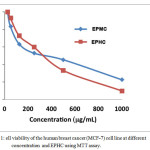
Figure 1 shows the percentage of cell viabilityusing MTT assay and the half maximal inhibitory concentration (IC50) of EPMC and EPHC against the human breast cancer (MCF-7)cell line. The IC50 value the concentration of the sample the cell viability was at 50%. Based on resultin Figure 1, the IC50 of EPMC was 360μg/mL EPHC was 340 μg/mL.The ability of microorganisms modifnatural product into other compounds activity compared to parental compound has attracted a great attention in recent years. biotransformed products screened for their activity and compared the parental compound.MTT was carried out to the cytoxicity of ethyl p-methoxycinnamate (EPMC) and product ethyl p-hydroxycinnamate (EPHC) against MCF-7 human breast cancer cell line. The assay was carried out to the minimum concentration of compound that could inhibit cell growth or in this case cause cell viability to decrease.p-hydroxycinnamate (EPHC) cytotoxicsince it lowe percentage of cell viability 9.87 % compared to ethyl p-methoxycinnamate (EPMC), was 22.58 % at the highest concentration (1000 μg/ml). However, both compoundcytotoxicMCF- line at all concentration used in assay.
 |
Figure 1: Ell viability of the human breast cancer (MCF-7) cell line at different concentration and EPHC using MTT assay. |
The half maximal inhibitory concentration (IC50) which is the measure of the effectiveness of a compound in inhibiting biological or biochemical function was . The readings were taken by measuring the concentration of the sample when the cell viability was at 50%. By result obtained in Figure 1, it can be said thatEPHCis more cytotoxic than EPMCas its IC50 value against MCF-7 was 340μg/mLwhile the IC50 of was 360μg/mL Therefore, from the result obtained, it can be concluded that Ethyl p-hydroxycinnamate (EPHC)potent since concentration to inhibit at least 50% growth of MCF-7 cancer cell line.
Conclusion
oth compounds (EPMC and EPHC)were active and exhibited good inhibition potential against MCF-7 cell lines. thyl p-hydroxycinnamate (EPHC)lower cell viability at high concentration 1000μg/mL compared to ethyl p-methoxycinnamate (EPMC). The IC50 value of EPHC was 340μg/mL against MCF-7 cell line. Therefore, the results of cytotoxicity studies and the IC50 values demonstrate potent selective toxicity property of ethyl p-hydroxycinnamate against the breast cancer cell line.In conclusion, thebiotransformed product, ethyl p-hydroxycinnamate (EPHC) has good potential asanti-cancer agent positive resultcompared to its parental compound, ethyl p-methoxycinnamate.
Acknowledgement
The authors would like to express greatest appreciation and gratitude to the International Islamic University Malaysia (IIUM) for financial support (RMGS).
References
- Omar, M. N.;M. Hasali, N. H. M.;Khan, N. T.; Moin, S. F.;AlFarra, H. Y.Biomedical & Pharmacology Journal2012, 5, 19-24.
- Omar, M. N.; Yusoff, N. S. A. M.; Zainuddin, N. A.; Zuberdi, A. M. Orient.J. Chem.,2014, 30, 1133-1136.
- Omar, M. N.; Shaban, N.; Bakar, L. M.; Zuberdi, A. M.Orient.J. Chem.,2014, 30, 1147-1151.
- Chen, G.; Chen, J. A. Appl. Microbiol. Biotechnol.2013, 97, 4325-4232, 2013.
- Omar, M. N.; AlFarra, H. Y.; Ichwan, S. J. A. Journal of Sustainable Science and Management 2016, in press.
- Omar, M. N.; Hasali, N. H. M.; AlFarra, H. Y.;Yarmo, M. A.; Zuberdi, A. M. Orient.J. Chem.,2014, 30, 1037-1043.
- Hasali, N.H.M.; Omar, M.N.;Zuberdi, A.M.; AlFarra, H. Y. International Journal of Biosciences2013, 3, 148-155.
- De Pooter, H. L.; Omar, M. .N.;Coolseat, B. A.; Schamp, N. M. Phytochemistry1985, 24, 93-96.
- Omar, M .N.; Razman, S.; Nor-Nazuha, M. N.;Nazreen, M. N. M.; Zuberdi, A .M. Orient.J. Chem.,2013, 29, 89-92.
- Omar, M. N. Journal of Tropical Agriculture and Food Science1991, 1, 147-152.
- Omar, M. N.; Ichwan, S. J. A.; Hasali, N. H. M.; Rahman, S. M. M. A.; Rasid, F. A.; Zuberdi, A. M. Jurnal Teknologi 2016. in press
- Kosuge, T.; Yokota, M.; Sugiyama, K.; Saito, M.; Iwata, Y.;Nakura, M.; Yamamoto, T. Chem. Pharm. Bull.1985, 33, 5565-5567.
- Zheng, G. Q.; Kenny, P. M.; Lam, L.K.T. J. Agric .Food. Chem.1993, 41, 153-156.
- Noro, T.; Miyase, T.; Kuroyanagi, M.; Ueno, A.; Fukushima, S. Chem. Pharm. Bull.1983, 31, 2708-2711.
- Jagadish, P.C.; Chandrasekhar, H.R.; Kumar, S.V.; Latha, K .P. Int. J. Pharm. Bio. Sci.2010,1, 1-5.
- Mosmann, T. Journal of Immunological Methods1983, 65, 55-63.

This work is licensed under a Creative Commons Attribution 4.0 International License.


