Virtual Screening Against Mycobacterium tuberculosisLipoate Protein Ligase B (MtbLipB)and In SilicoADMETEvaluation of Top Hits
Junie B. Billones1,2*, Maria Constancia O. Carrillo1, Voltaire G. Organo1, Stephani Joy Y. Macalino1, Inno A. Emnacen1, Jamie Bernadette A. Sy1
1OVPAA-EIDR Program: Computer-aided Discovery of Compounds for the Treatment of Tuberculosis in the Philippines, Department of Physical Sciences and Mathematics, College of Arts and Sciences 2Institute of Pharmaceutical Sciences, National Institutes of Health University of the Philippines Manila Taft Avenue, Ermita, Manila, Philippines 1000
DOI : http://dx.doi.org/10.13005/ojc/290423
Article Received on :
Article Accepted on :
Article Published : 14 Jan 2014
The emergence of drug resistant strains of Mycobacterium tuberculosis(Mtb) has spurred the search for new therapeutic targets for the development of more efficient anti-tuberculosis drugs. Lipoate protein ligase B (LipB), an enzyme involved in the biosynthesis of the lipoic acid cofactor, is considered as a very promising drug target in M. tuberculosis, since the bacteria has no known substitute enzyme that can take over the role of LipB in its metabolic system. Hence, apharmacophore-based screening, docking, and ADMET evaluation of compounds obtained from the National Cancer Institute (NCI) Database were performed against the MtbLipB enzyme. Consequently,nine compounds with superior binding energies compared to its known inhibitor (decanoic acid) have been identified. Moreover, among these nine compounds, NSC164080 (methyl 2-(2-(((benzyloxy)carbonyl)amino)propanamido)-3-(4-hydroxyphenyl)propanoate) displayed the most favorable ADMETproperties. The results in this work may pave the way for the development of a novel class of antituberculosis agents.
KEYWORDS:ADMET;antituberculosis compound; lipoate protein ligase (LipB); Mycobacterium tuberculosis; octanoyl-[acyl carrier protein]-protein acyltransferase; pharmacophore; virtual screening
Download this article as:| Copy the following to cite this article: Junie B. B, Maria C. O. C, Voltaire G. O, Stephani J. Y. M, Inno A. E, Jamie B. A. S. Virtual Screening Against Mycobacterium tuberculosisLipoate Protein Ligase B (MtbLipB)and In SilicoADMETEvaluation of Top Hits. Orient J Chem 2013;29(4) |
| Copy the following to cite this URL: Junie B. B, Maria C. O. C, Voltaire G. O, Stephani J. Y. M, Inno A. E, Jamie B. A. S. Virtual Screening Against Mycobacterium tuberculosisLipoate Protein Ligase B (MtbLipB)and In SilicoADMETEvaluation of Top Hits. Orient J Chem 2013;29(4). Available from: http://www.orientjchem.org/?p=1599 |
INTRODUCTION
Tuberculosis (TB) is an infectious disease caused by Mycobacterium tuberculosis that affects the lungs. At present, TB remains to be one of the most prevalent infections, resulting in almost 1.4 million deaths and an estimated number of 8.7 million new TB cases worldwide in 2011 [1]. Approximately two billion people or about a third of the world’s population is infected with TB, most of which having latent TB [2]. TB is the sixth biggest cause of death in the Philippines; making it as onewith the highest TB incidences in Asia [3]. Although drugs such as isoniazid and rifampicin are available to treat TB, the emergence of multi-drug resistant (MDR) and extensively-drug resistant (XDR) tuberculosis poses a big challenge to the treatment of tuberculosis [4]. The alarming rise of resistant strains of Mycobacterium tuberculosis against current drugs calls for an increase in research efforts towards the development of new therapeuticsagainst diverse strains of M. tuberculosis.
The enzymes involved in lipoylation and lipoic acid, which is essential for the activation of several protein complexes participating in key metabolic processes, have been implicated in the growth and pathogenicity of some bacteria including Mycobacterium tuberculosis. Lipoate protein ligase B (LipB), also known as octanoyl-[acyl carrier protein]-protein acyltransferase, is
an enzyme that catalyzes the transfer of endogenous octanoic acid to lipoyl domains by way of a thioester bond to the 4’-phosphopanthetheine cofactor of the acyl carrier protein (ACP) [5,6]. An S-adenosyl-L-methionine-dependent enzyme known as lipoyl synthase (LipA) then converts the octanoyl derivatives into lipoyl derivatives by catalyzing the insertion of sulfur atoms into the six- and eight-carbon positions of the resulting fatty acid. This process circumvents the need of the bacteria for exogenous lipoic acid [5]. Expression of LipB was found to be significantly up-regulated in patients with multi-drug resistant M. tuberculosis and has no known back-up mechanism that can take over its role in the metabolism of TB [7]. Moreover, previous efforts to generate a knockout model lacking the lipB gene has resulted in consistent failure [5], supporting the theory that LipB is essential in the growth of Mycobacterium tuberculosis, making it a viable target for the search of novel anti-TB drugs.
In vitro screening of candidate drugs against TB has been done for several years with varying rates of success. However, wet laboratory approaches have been hampered by serious limitations such as the requirement of a BSL3 laboratory equipped with high-throughput equipment and materials, which entails the need for a great amount of money and expert research skills in handling the arduous and sensitive protocols used in drug screening [8].
Nevertheless, computational methods have recently been gaining popularity as it can be used for molecular docking, molecular simulations, and de novo design, as well as ligand- or structure-based virtual screening of drug candidates. These techniques are able to evaluate binding affinity of a ligand to a receptor to gauge its pharmacological activity, which is helpful in prioritizing candidate compounds for synthesis and screening in the laboratory [9]. In particular, structure-based virtual screening simply requires 3D structural data of both the protein and the drug candidates, which are made available by laboratory procedures such as X-ray crystallography and NMR Spectroscopy.In this work, a pharmacophore derived from the structure of LipBwas used to virtually screena database of compounds for new leadsagainst Mycobacterium tuberculosis. Moreover, the high-scoring compounds were further sieved by the use of in silico ADMET filters.
MATERIALS & METHODS
Structural data of LipB protein and compound library. The 1.08Å – resolution3D structure of LipBcomplexed with decanoic acid (PDB ID: 1W66) was retrieved from the Protein Database (www.rcsb.org). The bound decanoic acid was removed and the protein was prepared using the Prepare Protein protocol of Discovery Studio 3.5 (Accelrys, Inc.) using the default parameters. After preparation, the prepared protein structure was minimized using the Minimization protocol using the default parameters. The root-mean-square deviation (RMSD) of the prepared protein was calculated using the Superimpose Proteins Tool.
The NCI database was downloaded from the National Cancer Institute download page (http://cactus.nci.nih.gov/download/nci/). The compounds in the NCI database were prepared using Prepare Ligands protocol. All parameter values used were default except for the Lipinski Filter, which was turned off.
Generation of Structure-based Pharmacophore Model.The pharmacophore was generated using the chemical features of the binding site of LipB. The location of the bound decanoic acid was chosen as the binding site of LipB. Further evidence to the identification of the binding site is the presence of Cys176, a strictly conserved residue, to which the decanoic acid was covalently attached by means of a thioester bond. The binding site sphere was defined using the Binding Site Tool in DS 3.5 with a radius of 12 Å, centered on the position of the bound decanoic acid. The Interaction Generation protocol of DS3.5 was used to generate the pharmacophore model containing the features (hydrophobic, H-donor, H-acceptor) for interaction in the protein’s active site. For screening, a maximum of 30 features are allowed, thus the Edit and Cluster PharmacophoresTool was used to cluster and edit the pharmacophore model down to <30 features.
Screening of Compound Libraries.A total of 153,000 (NSC1 to NSC257903) compounds from NCI database were screened in this work. The Prepare Ligands protocol generated several conformations per compound in the libraries, all of which were used in the screening process. After preparation, the Build 3D Database protocol was used to create compound databases for
easier screening. The generated structure-based pharmacophore model was used to screen the compound databases using the Screen Library protocol, which employs the flexible search method of Catalyst. Screening was done twice, one for rigid fitting method and another for flexible fitting method. The hit compounds were selected based on their fit values.
Molecular Docking. The hit compounds from the pharmacophore screening were docked to the LipB active site using the CDOCKER docking protocol. Decanoic acid was also docked and compared to the original bound conformation to validate the docking method.The calculated binding energy was employed as the baseline comparison for the selection of compounds with the best binding affinity to LipB. Calculate Binding Energies protocol was used to compute for the binding affinity of the hit compounds. All compounds with better binding affinity than decanoic acid were selected for further screening.
In silicoADMET. The compounds with remarkably high binding affinities were further screened using ADMET filters. Accordingly, the solubility, absorption, plasma protein binding, CYP2D6 inhibition, and hepatotoxicitywere determined using the ADMETand TOPKAT protocols of DS 3.5.
RESULTS AND DISCUSSION
The Prepare Protein protocol,which was used for the LipB (PDB ID: 1W66) (Figure 1A) preparation, primes the protein for input into other protocols in DS3.5 by inserting missing atoms in incomplete residues, optimizing side-chain conformation, modeling missing loop regions, removing alternate conformations, and protonating titratable residues [10]. Minimization was performed on the prepared protein structure to find the most stable protein conformation [10]. This procedure marginally changed the original conformation of the protein, thus, RMSD value was calculatedin order to measure the change in conformation during geometry optimization. Superimposition of the two protein conformations revealed only minor deviation (RMSD = 0.71 Å) of the docked structure from the native conformation (Figure 1B).
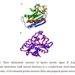 |
Figure 1.(A) Three dimensional structure of lipoate protein ligase B (LipB) [5].The Mycobacterium tuberculosis LipB enzyme functions as a cysteine/lysine dyad transferase.), (B) Molecular overlay of downloaded protein structure (blue) and prepared protein structure (pink). Click here to View figure |
The Prepare Ligands protocol prepares the ligand by removing duplicate structures, generating isomers and tautomers, generating 3D conformations and other tasks established by user-defined parameters [10]. The protocol also includes a Lipinski filter, which employs the Lipinski’s rule of five for initial screening of ligands. Lipinski showed that small-molecule oral drugs follow a set of rules dubbed as the ‘rule of five’: MW< 500 Da; LogP< 5; H-bonds< 5, and donors and acceptors are less than or equal to 5[11]. Any compound thatviolates these rules is less likely to be absorbed orally. However, several studies have already showed that some compounds, including many natural products and natural product-like molecules, regardless of being outliers to the criteria set by the Lipinski rule, have become successful candidate drugs [12]. Thus, the Lipinski filter was not employed in the screening process.
A pharmacophore is a set of steric and electronic features used to screen compounds to help prioritize compounds with optimal ligand interactions with the biological target. Pharmacophore features specify areas in a molecule that are hydrophobic, hydrogen donor or hydrogen acceptor
[10]. Before generation of the pharmacophore, the binding site was defined based on the approximate position of the bound decanoic acid (i.e. 9.81, 10.01, 5.81), and the radius was set at 12 Å (Figure 2A). The pharmacophore was generated using the chemical features of the binding site of LipB showing the hydrophobic, H-donor, and H-acceptor features in 3D space (Figure 4B).
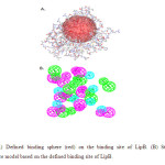 |
Figure 2. (A) Defined binding sphere (red) on the binding site of LipB. (B) Structure-based pharmacophore model based on the defined binding site of LipB. Click here to View figure |
After generating the pharmacophore model, virtual screening of the ligand database was executed, and the compounds were rank-ordered according to their fit values.Rigid and flexible screening processes were performed in succession.In particular, after rigid screening, only those compounds with fit valuesgreater than 2.5 were carried out for subsequent flexible screening process. In the flexible fitting method, each ligand conformation was slightly modified to better fit the pharmacophore model [10].
The arbitrary cut-off fit value of 3.0 was used as basis for further reduction of compounds down to a manageable number. Accordingly, the high-scoring compounds listed in Table 1, were then subjected to molecular docking calculations.
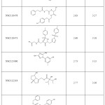 |
Table 1. Rigid and flexible screening fit values of the hit compounds from NCI database. Click here to View table |
The hit compounds (Flexible Screening fit value > 3.0), as well as decanoic acid were subsequently docked to the defined binding site of LipB using CDOCKER docking protocol in DS3.5. CDOCKER (CHARMm-basedDOCKER) is a grid-based molecular dynamics (MD) simulated-annealing-based algorithm docking method that employs CHARMm force fields and allows for full ligand flexibility in docking process. It generates several ligand poses when the ligand is docked into the receptor’s binding site and implements molecular dynamics-based simulated annealing and in situ minimization [10]. The binding affinity of each hit compound was computed using the Calculate Binding Energiesprotocol, and evaluated against the binding energy of decanoic acid. In this protocol, each pose of a compound was allowed to minimize in situ to determine the best interaction between the ligand and the residues within the binding site of the receptor.
Decanoic acid, the known inhibitor of LipB[6], was docked to the receptor to validate the docking procedure. Figure 3C showed that the docked decanoic acidformed most of the interactions that were observed experimentally (Figure 3B)such as van der Waals interaction with Gly78, Gly147, Ala145, Ala160, Phe159, Thr81, His83, Arg76, and Ile146. It is noteworthy thatthe docked decanoic acid formed polar interactions with Cys176 in accord with experiment, albeit thioester covalent formation was actually observed. Moreover,the ligand interacted with Lys79 through main chain H-bonding and polar interaction as well.Gratifyingly, the calculatedRMSD value of 1.03 Å for theoverlayedligand conformations (Figure 3A)further demonstrates the validity of our docking protocol.
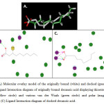 |
Figure 3. (A) Molecular overlay model of the originally bound (white) and docked (green) decanoic acid. (B) Ligand Interaction diagram of originally bound decanoic acid displaying thioester bond with Cys176 (yellow circle) and various van der Waals (green circle) and polar (magenta circle) interactions. (C) Ligand Interaction diagram of docked decanoic acid. Click here to View figure |
The molecular docking study furnishednine compounds from the NCI database with greater binding affinity to LipBcompared to decanoic acid (Table 2). Interestingly, the top 2 compounds (NSC211851 and NSC245342) are polyols while the rest contain a combination of functionalities including amines, amides, ester, carboxylic acid, and alcohol. Furthermore, the ligand interaction diagrams were generated to determine the component factors that contributed to the overall binding potential, that is, the number of van der Waals, polar, H-bond, and pi interactions that the high affinity compounds formed with the residues found in the active site of LipB (Table 3).Evidently, most of the compounds formed either polar interaction or H-bonding with Lys79 and Cys176, which are distinctly featured in the crystal structure of the LipB complex.
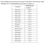 |
Table 2. Binding energy for the best pose (docked conformation) of the top hitsin complex withLipBby the use of CDOCKER protocol in Discovery Studio 3.5. Click here to View table |
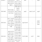 |
Table 3. Ligand interactions (van der Waals, polar, hydrogen bond, and pi interaction) of bound and docked decanoic acid and the hit compounds with the best binding affinities. Click here to View table |
The top nine compounds from the molecular docking study were subsequently subjected to final screening using ADMET and TOPKAT protocols in DS3.5 to predict their ADMET properties.
Among the nine compounds, NSC164080 (methyl 2-(2-(((benzyloxy)carbonyl)amino)propanamido)-3-(4-hydroxyphenyl)propanoate), a compound containing ester, amido, and phenolic functionalities, had the most favorable ADMET properties and showed the least toxicity (vide infra) (Table 4). Interestingly, in addition to numerous van der Waals, polar and pi interactions, NSC164080 displayed side-chain hydrogen bond interactions with Cys176 and Arg58 (Figure 4). As pointed out above, a strong interaction with Cys176 is a hallmark of LipB inhibition.
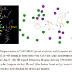 |
Figure 4. (A) 3D representation of NSC164080 (green) interaction with keyamino acid residues at the binding site. NSC164080 formed pi interactions with His83 and Arg58 and intermolecular H-bonding with Cys176 and Arg58. (B) 2D Ligand Interaction Diagram showing NSC164080 van der Waals (green circles), polar (magenta circles), H-bond (blue broken lines) and pi interaction (orange line) with amino acid residues in the binding site of the LipB receptor. Click here to View figure |
Traditionally, drugs are discovered in the laboratory using High-Throughput Screening (HTS) and several in vitro and in vivo biological tests, after which tests for pharmacokinetic and toxicity properties are performed. Usually, adverse findings are revealed at this late stage of drug discovery and development [13]. Fortunately, in silicotoxicity techniques such as predictive quantitative structure-activity relationship (QSAR), predictive ADMET, and other computational toxicity applications are now available for a priori assessment of pharmacokinetic properties of a compound [14]. The use of in silico toxicity and ADMET predictions pre-empts the need for wet laboratory toxicity testing and helps save time and effort in drug discovery ventures.
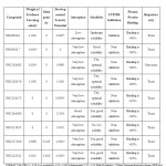 |
Table 4. ADME-Toxicity information on the hit compounds with the best binding affinities. Click here to View table |
Hence, The ADMET and TOPKAT predictive toxicity protocols in DS3.5 were employed to gather theADMET information of the top hit compounds. As shown in Table 4, most of the compounds indicate either carcinogenicity or toxicity potential. It is worthy of note, however, that the strongest
binding compound (NSC211851) also appeared to be the least toxic (i.e. non-carcinogenic and non-mutagenic with hardly any developmental toxicity potential). Unfortunately, being a polyol, it has very low absorption ability and very high solubility,which would effect undue excretion from the body. Fortuitously, there exists a compound (NSC164080),which indicates more favorable ADMET properties.NSC164080 has also very low probability of being carcinogenic, mutagenic, and toxic in an animal model. But in contrast to NSC211851 it promises to have good intestinal absorption ability and solubility, and does not inhibit CYP2D6, which can help in metabolizing and in flushing out the drug from the body. The only apparent drawbacks of NSC164080 are its tendency to be hepatotoxic and its over 90%plasma protein binding (PPB) potential. Nevertheless, the hepatotoxicity and the PPB of the compound may be alleviated in a dose-dependent manner such thatthe distribution of the drug in the liver and its circulation do not reach the critical levels [15, 16]. In fact, there are several hepatotoxic drugs that are out in the market such as acetaminophenthat are administered to patients in a strict dosage regimen [15]. Finally, these potential lead compounds can be further tweaked structurally to improve their potency and minimize their toxicity.
CONCLUSION
In conclusion, virtual screening and molecular docking of compounds from NCI database were performed to identify existing compoundswith high binding affinity with the MtbLipBenzyme target. Out of the 153,000 compounds, 9 showed superior binding energies than that ofthe known inhibitor, decanoic acid. Further ADMET calculations revealed that compound NSC164080 is a promising lead compound due to its satisfactoryADMET properties. NSC164080 exhibits a favorable side-chain hydrogen bond interaction with Arg58 and especially Cys176, which is featured in the crystal structure of decanoic-LipB complex. These results are certainly enlightening and helpful in future anti-TB drug discovery efforts. In fact, in vitro testing and lead optimization studies are underway in our group.
CONFLICT OF INTEREST
There is no conflict of interest.
ACKNOWLEDGEMENT
This study is generously supported by the Office of the Vice President for Academic Affairs (OVPAA), University of the Philippines System under the Emerging Inter-Disciplinary Research (EIDR) program (OVPAA-EIDR 12-001-121102).
REFERENCES
- WHO (2012). World Health Organization Global Tuberculosis Report 2012. http://www.who.int/tb/publications/factsheet_global.pdf.(Accessed March 2013)
- Frieden, T. R.; Sterling, T. R.; Munsiff, S. S.; Watt, C. J.; Cye, C.Tuberculosis. The Lancet,2003, 887–899.
- Department of Health.Leading causes of mortality. http://www.doh.gov.ph/node/198.html (Accessed March 2013)
- Sharma, S.K.; Mohan, A. Multidrug-resistant tuberculosis. Indian J. Med. Res.,2004, 120, 354 – 376.
- Ma, Q.; Zhao, X.;Eddine, A.; Geerlof, A.; Li, X.; Cronan, J.E. The Mycobacterium tuberculosis LipB enzyme functions as a cysteine/lysine dyad transferase. P. Natl. Acad. Sci. USA,2006,103(23): 8662 – 8667.
- Spalding, M.D.;Prigge, S.T. Lipoic acid metabolism in microbial pathogens.Microbiol. Mol. Biol. R.,2010, 74(2), 200–228.
- Rawal, S.;Sood, R.;Mahajan, N.; Sharma, M.; Sharma, A., Current status and future prospects for tuberculosis. Int.J.Pharm.Sci.Res.,2010,1(5), 128 – 134.
- Stanley, S.A.; Grant, S.; Kawate, T.; Iwase, N.; Shimizu M. Identification of novel inhibitors of M. tuberculosis growth using whole cell based high-throughput screening.ACS Chem. Biol.,2012, 7, 1377–1384.
- Lounnas, V.; Ritschel, T.; Kelder, J.; McGuire, R.; Bywater, R.P.; Foloppe, N. Current progress in structure-based rational drug design marks a new mindset in drug discovery. Comp. Struc. Biotech. J., 2013, 5(6), e201302011.
- Discovery Studio 3.5 Tutorials. Theory – Pharmacophore.AccelrysInc, San Diego, CA, USA,2012.
- Lipinski, C.A.; Lombardo, F., Dominy, B. W.; Feeney, P. J.Experimental and computational approaches to estimate solubility and permeability in drug discovery and development settings.Adv. Drug Deliver. Rev., 2001,46, 3–26.
- Murray, C.W.; Rees, D.C., The rise of fragment-based drug discovery. Nat. Chem.,2009, 1, 187– 192.
- van de Waterbeemd, H.; Gifford, E. ADMET in silico modelling: towards prediction paradise? Nature, 2003, 2, 192–204.
- Valerio, L. G.Jr. ,In silico toxicology for the pharmaceutical sciences. Toxicol. Appl. Pharm. 2009, 356–370.
- Lee, W.M. Drug-Induced Hepatotoxicity. The New England J. Med.,2003, 474–485.
- Balakin, K.V.; Ivanenkov, Y.A.; Sabchuk, N.P.; Ivashchenko, A.A.; Ekins, S. Comprehensive computational assessment of ADME properties using mapping techniques. Curr. Drug Discov. Tech.,2005, 2, 99–113.

This work is licensed under a Creative Commons Attribution 4.0 International License.


